Email
:
cs@trivedieffect.com
    trivedi-physiology-logo-1
    trivedi-physiology-logo
    trivedi-physiology-logo-1
    • OVERVIEW
    • CIRCULATORY
      SYSTEMS
      • ARTERIAL PULSE WAVE ANALYSIS
      • TREADMILL TEST REPORT
      • ECHO CARDIOGRAPHY REPORT
      • HEMATOLOGY REPORT
    • CRANIOSACRAL
      SYSTEMS
      • DR. LORI CROTEAU'S REPORT
      • DEBBIE POPE’S CRANIAL SACRAL REPORT
      • DR. DEREK A. PYPER'S REPORT
    • DIGESTIVE
      SYSTEMS
      • ULTRASONOGRAPHY(USG) ABDOMEN WITH PELVIS REPORT
    • ENDOCRINE
      SYSTEMS
      • UNIQUE PHYSIOLOGY OF PITUITARY GLAND (DR. PATIL‘S REPORT)
      • BRAIN MAGNETIC RESONANCE IMAGING (MRI) IMAGES
      • SKULL X-RAY IMAGES
      • HORMONE REPORT – 2008
      • HORMONE REPORT – 2007
      • BIOCHEMISTRY REPORT
    • MUSCULOSKELETAL
      SYSTEMS
      • ORTHOPEDIC REPORT
      • X- RAYS IMAGES
      • SPINE MAGNETIC RESONANCE IMAGING (MRI) IMAGES
      • DR RICO PROVASOLI‘S SPINE REPORT
      • CHIROPRACTIC EVALUATION IN 2012 BY DR. STEVE HRUBY
      • CHIROPRACTIC AND NEUROLOGICAL REPORT, DR, HRUBY, 2013
    • NERVOUS
      SYSTEM
      • COMPUTERIZED BIOFEEDBACK REPORT
      • MEDICAL INFRARED DIGITAL IMAGING (MIDI)
      • ELECTROMYOGRAPHY (EMG) REPORT
      • ELECTROENCEPHALOGRAPHY (EEG) REPORT
    • RESPIRATORY
      SYSTEMS
      • PULMONARY FUNCTION TEST (PFT) REPORT
    Hit enter to search or ESC to close

    CHIROPRACTIC EVALUATION IN 2012 BY DR. STEVE HRUBY

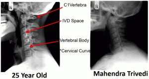
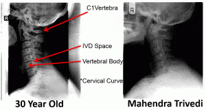
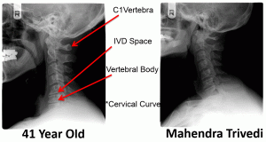
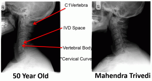
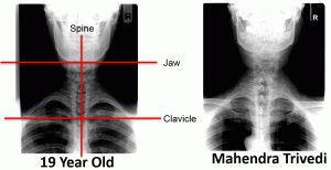
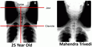
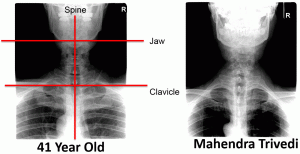
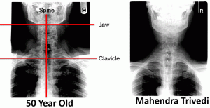
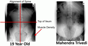
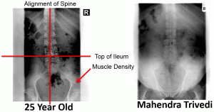
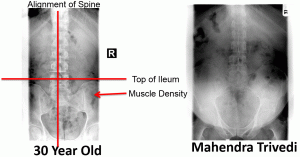
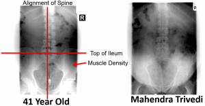
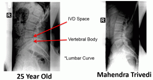
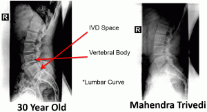
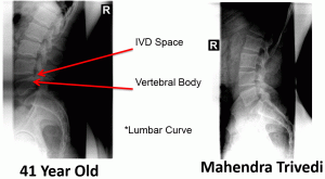
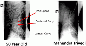
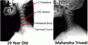
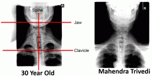
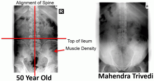
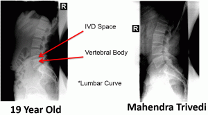
    On November 8th, 2012 in Scottsdale, Arizona, Dr. Steve Hruby, D.C. performed a chiropractic evaluation on Mahendra Trivedi. The following are Spinal X-rays comparing Mr. Trivedi’s (age 49) results with X-rays of males ages 19-50.

    The results are miraculous.
    Chiropractic Evaluation

    • Spine
      • Inner vertebral discs
      • Degeneration
      • Shape, gaps, cushion
    • Bones
      • Density
      • Curvature of spine
    • Posture
    • Pelvic area
    • Muscles
      • Density
      • Flexibility
      • Tonicity
    • Joints
      • Active and passive range of motion
      • Central nervous system
    • Diaphragm
     

    Technologies Used During Evaluation

    • Digital X-ray
    • SEMG
    • Thermography
    • Palpation
    • Active Range of Motion (AROM)
    • Postural Evaluation

    Comparison of Lateral Cervical Spine
    1 - steve1-300x156
    2 - steve2-300x160
    3 - steve3-300x160
    4 - steve4-300x160-2
    5 - steve5-300x160-1

    Comparison of AP Cervical Spine
    6 - steve6-300x154
    7 - steve7-300x153
    8 - steve8-300x151
    9 - steve9-300x154
    10 - steve10-300x154

    Comparison of AP Lumbo Pelvic
    11 - steve11-300x158
    12 - steve12-300x158
    13 - steve13-300x157
    14 - steve14-300x156
    15 - steve15-300x159

    Comparison of lateral lumbo Spine
    16 - steve16-300x166-1
    17 - steve17-300x156-1
    18 - steve18-300x163-1
    19 - steve19-300x165-1
    20 - steve20-300x163-1

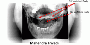
    Ap open mouth

    21 - steve21-300x150

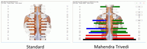
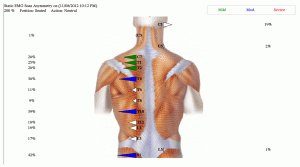
    Surface Electromyography (EMG)

    22 - steve22-300x100

    Detects the electrical potential generated by muscle cells when these cells are electrically or neurologically activated. Essentially this technology is used to assess the level of communication between the brain and the body.

    Surface EMG (Muscle Symmetry)
    23 - steve23-300x167-1
    Summary of Exam Results
    • Spine comparable to that of a healthy 19 year old
    • Little to no degeneration of cervical and lumbar spine (highly abnormal for a 49 year old)
    • Ideal position of 1st cervical vertebrae
    • Ideal alignment of both cervical and lumbar spine
    • Normal lordosis in cervical and lumbar spine (forward curve of spine), (highly abnormal)
    • Abnormal density (high density) of pelvic muscles, not usually viewed in x-rays
    • Ideal alignment of pelvic bones on evaluation and X-rays
    • Ideal range of motion of cervical spine
    • Thick supple muscles in cervical and lumbar spine (highly abnormal)
    Copyright 2025. All Rights Reserved by Trivedi Physiology | Home